What anatomical structures must be
avoided during the placement of
dental implant?
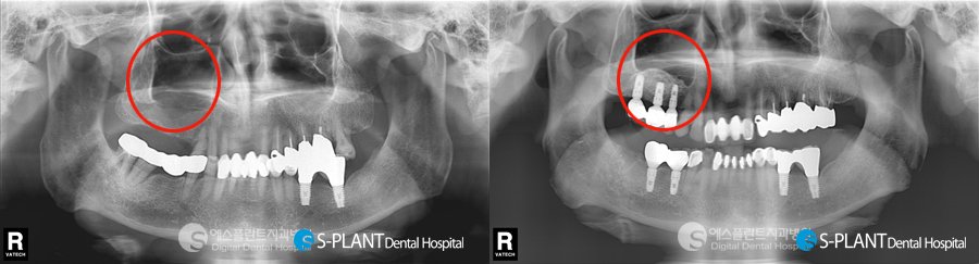
For Upper Jaw
In the upper jaw, provided the implants
stay within the bone that once supported
your own teeth there are really no
important risk areas. If you have missing
upper back teeth then the shape and
location of the region above the roots
(maxillary sinus) can be shown to you.
The maxillary sinuses can be seen on most
x-rays and are therefore readily avoided.
For Lower Jaw
In the lower jaw the most important
anatomical structure to be avoided is t
he ‘inferior dental nerve’.
This nerve runs from the area behind
the wisdom teeth, passes under the back
teeth(molars) and emerges onto the skin
of the face in the region where your
middle teeth (premolars) are or used to be.
This is why a normal dental anaesthetic
produces a numb lip even when the needle
was placed right at the back of the mouth.
If this nerve is disturbed or damaged during
the placement of dental implants it can lead
to temporary or even permanent numbness
or altered sensation. This is a rare but
important complication.
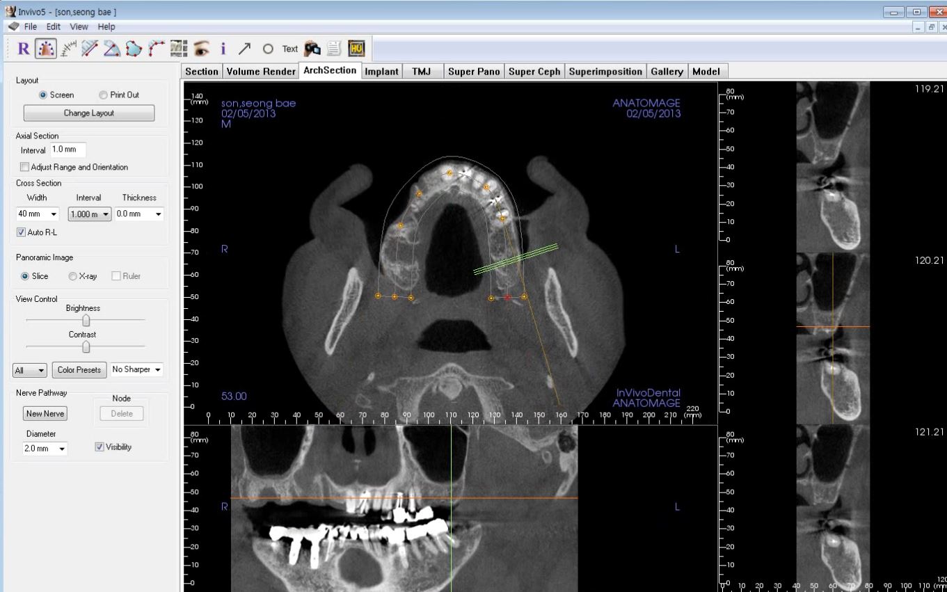
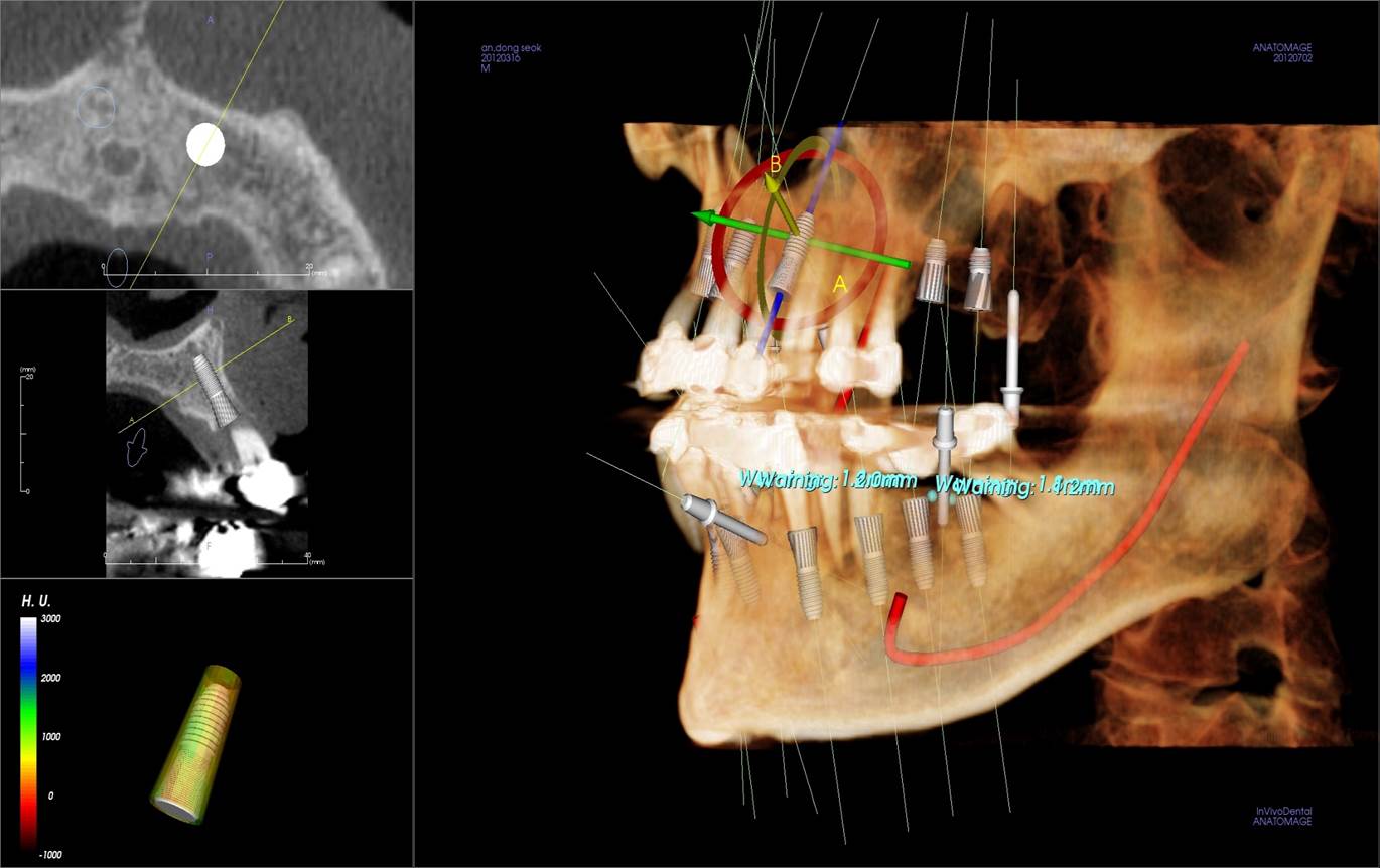
S-PLANT 3D CT scans are generally
the best means for identifying the location
of this nerve and allow implants to be
placed with considerable confidence;
however, these are only sometimes
available within a normal dental surgery
environment. It may therefore require
a visit to a suitable hospital
where the scan is generally completed
within a few minutes. Whilst CBCT
scans are more expensive than routine
dental x-rays, the information
they provide is often invaluable for
complex treatment planning and
knowing where important anatomical
structures are located.

'English > Implant' 카테고리의 다른 글
| Could missing teeth cause dementia? (0) | 2019.05.25 |
|---|---|
| Dental Implants for Diabetic Patients (0) | 2019.05.13 |
| 3D virtual implants planning and flapless implant surgery using surgical guide. (0) | 2019.04.16 |
| How long do dental implant last? (0) | 2019.04.09 |
| ‘Bone’ - The foundation for dental implants (0) | 2019.04.02 |


